Pellucid Marginal Degeneration and Specialty Contact Lenses in Red Bank, NJ
Pellucid marginal degeneration, often shortened to PMD, is a rare corneal thinning condition that can make vision blurry, distorted, or difficult to correct with standard glasses. Because PMD can resemble keratoconus and other corneal shape changes, careful evaluation is important.
At Crystal Eyecare in Red Bank, NJ, patients with irregular corneas may benefit from a detailed eye exam and a discussion about specialty contact lens options. These lenses are designed to help manage complex vision needs when traditional soft contacts or glasses may not provide the clearest correction.
For patients who have been told they have PMD, keratoconus, irregular astigmatism, or another corneal ectasia, custom contact lens fittings may be an important part of the vision care plan.
What Is Pellucid Marginal Degeneration?
Pellucid marginal degeneration is a non-inflammatory corneal ectasia. In plain English, the cornea gradually thins and changes shape. The cornea may stay clear, but its irregular shape can bend light unevenly and create distorted vision.
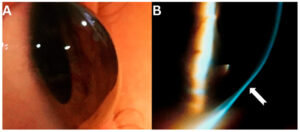
PMD most often involves thinning in the lower outer portion of the cornea. This can lead to high or irregular astigmatism. Some people notice frequent prescription changes, glare, ghosting, double images from one eye, or difficulty seeing clearly at night.
PMD is usually monitored over time because progression can vary. Some cases are mild and stable, while others may create more significant vision challenges.
How PMD Can Affect Vision
The main issue with PMD is not only corneal thinning. It is the irregular optical shape created by the thinning and bulging pattern. When the cornea becomes irregular, glasses may not fully sharpen vision because glasses sit in front of the eye and cannot reshape the corneal surface.
Common visual symptoms can include:
- Blurred or distorted vision
- Ghosting or shadow images
- Glare or halos, especially at night
- Frequent changes in glasses prescription
- Reduced clarity even with updated glasses
- Difficulty with standard soft contact lenses
These symptoms can also occur with other eye conditions, so an eye exam is needed to understand the cause.
PMD Versus Keratoconus
PMD and keratoconus are both corneal ectatic conditions. Both can thin and distort the cornea, and both can cause irregular astigmatism. However, the location and pattern of thinning are usually different.
Keratoconus often involves a more central or lower-central cone-shaped steepening of the cornea. PMD classically involves a lower peripheral band of thinning, with the area of bulging often sitting just above that thinned zone.
A corneal topography pattern sometimes called a “crab claw” or “kissing doves” pattern can suggest PMD, but it is not enough by itself to confirm the diagnosis. Inferior keratoconus can sometimes look similar on topography. This is why eye doctors often use a combination of slit-lamp exam findings, corneal topography or tomography, corneal thickness mapping, and follow-up measurements.
How Eye Doctors Evaluate PMD
A PMD evaluation may include several steps, depending on the patient’s history, symptoms, and exam findings. The goal is to understand the shape, thickness, and stability of the cornea.
Evaluation may include:
- A complete refraction to measure the glasses prescription
- Slit-lamp examination of the cornea
- Corneal topography or tomography
- Corneal thickness measurements
- Assessment of best-corrected vision
- Review of prior records, when available
- Monitoring for changes over time
This type of careful assessment helps distinguish PMD from keratoconus, Terrien marginal degeneration, post-surgical ectasia, and other corneal conditions.
Why Specialty Contact Lenses May Help
When the cornea is irregularly shaped, specialty contact lenses may help create a smoother optical surface. This can improve visual clarity for some patients with PMD, keratoconus, post-surgical corneas, and other complex corneal conditions.
Specialty lens options may include rigid gas permeable lenses, hybrid lenses, piggyback systems, and scleral lenses. The right option depends on corneal shape, comfort, vision goals, eye health, and handling ability.
Scleral lenses are often discussed for irregular corneas because they vault over the cornea and rest on the white part of the eye. This design can help reduce the effect of corneal irregularity on vision. Not every patient needs or qualifies for scleral lenses, but they can be an important option in the specialty contact lens toolkit.
Crystal Eyecare offers custom contact lens fittings for patients who need more than standard contact lenses. This may include patients with irregular astigmatism, keratoconus, PMD, dry eye challenges, or other complex fitting needs.
A Stepwise Approach to PMD Vision Care
PMD care is usually individualized. There is no one-size-fits-all plan. The best approach depends on how much the cornea has changed, whether the condition is progressing, and how well the patient sees with glasses or contact lenses.
In mild cases, glasses may provide acceptable vision. When the corneal shape becomes more irregular, standard glasses may become less effective. At that point, specialty contact lenses may be considered to improve the optical surface.
In cases where progression is documented, a corneal specialist may discuss treatments such as corneal collagen cross-linking. Surgical options are generally reserved for more advanced cases or for patients who cannot achieve functional vision with other approaches. Crystal Eyecare can help patients understand their options and coordinate care when specialist referral is appropriate.
PMD and Cataract Surgery Planning
PMD can also matter when cataract surgery is being planned. Because the cornea helps determine the focusing power of the eye, an irregular or changing corneal shape can make measurements more complex.
Patients with known or suspected PMD may need careful corneal measurements before cataract surgery. In some cases, contact lens wear may need to be paused before measurements are taken. Stable corneal findings are especially important when discussing astigmatism correction or premium lens options.
This is another reason regular eye care and clear communication between providers are important.
When to Schedule an Eye Exam
Schedule an eye exam if you have blurry vision that does not seem fully corrected with glasses, frequent prescription changes, ghosting, glare, or difficulty with standard contact lenses. These symptoms do not automatically mean you have PMD, but they are worth evaluating.
You should seek urgent or emergency care for sudden vision loss, severe eye pain, new marked redness, trauma, sudden corneal clouding, flashes, floaters, curtain-like vision loss, or neurologic symptoms such as weakness, numbness, speech difficulty, severe headache, or double vision with neurologic signs.
Specialty Contact Lens Care in Red Bank, NJ
For patients in Red Bank, Monmouth County, and nearby New Jersey communities, Crystal Eyecare provides a local option for specialty contact lens evaluation. A custom fitting is more detailed than a standard contact lens visit because the lens must be matched to the eye’s unique shape, visual needs, and comfort requirements.
If you have been told that you have PMD, keratoconus, irregular astigmatism, or a complex corneal shape, ask whether custom contact lens fittings may be appropriate for your situation. Typically, scleral lenses are warranted.
Call now — see what you have been missing.
Key Takeaway
Pellucid marginal degeneration is a rare corneal thinning condition that can make vision difficult to correct with standard glasses or contacts. A careful diagnosis matters because PMD can resemble keratoconus and other corneal conditions. For many patients with irregular corneal optics, specialty contact lenses may play an important role in improving day-to-day visual function.
Crystal Eyecare in Red Bank, NJ can help patients explore eye exam findings, specialty contact lens options, and next steps for coordinated care when needed.
References
- Sahu, J., & Raizada, K. Pellucid Marginal Corneal Degeneration. StatPearls, NCBI Bookshelf. https://www.ncbi.nlm.nih.gov/books/NBK562314/
- Tsatsos, M., et al. Pellucid Marginal Degeneration: A Comprehensive Review of Pathophysiology, Clinical Features, Diagnosis, and Management. Journal of Clinical Medicine, 2025. https://pubmed.ncbi.nlm.nih.gov/40806799/
- Vieira, I. V., et al. Update on Pellucid Marginal Degeneration. Graefe’s Archive for Clinical and Experimental Ophthalmology, 2025. https://pubmed.ncbi.nlm.nih.gov/41212223/
- Martínez-Abad, A., & Piñero, D. P. Pellucid Marginal Degeneration: Detection, Discrimination From Other Corneal Ectatic Disorders and Progression. Contact Lens & Anterior Eye, 2019. https://pubmed.ncbi.nlm.nih.gov/30473322/
- Koc, M., et al. Crab Claw Pattern on Corneal Topography: Pellucid Marginal Degeneration or Inferior Keratoconus? Eye, 2018. https://www.nature.com/articles/eye2017198
- Gruenauer-Kloevekorn, C., et al. Pellucid Marginal Corneal Degeneration: Evaluation of the Corneal Surface and Contact Lens Fitting. British Journal of Ophthalmology, 2006. https://pmc.ncbi.nlm.nih.gov/articles/PMC1856967/
- Rathi, V. M., et al. Scleral Contact Lenses in the Management of Pellucid Marginal Degeneration. Contact Lens & Anterior Eye, 2016. https://pubmed.ncbi.nlm.nih.gov/26669275/
This article is for educational purposes only and is not a diagnosis or a substitute for professional eye care. An eye exam is needed to evaluate your individual vision and eye health needs.