Hopefully a Helpful FAQ
FAQs
by Alvaro Cordova
After doing this a while, these are the most common questions I get. The eye picture should help when specific structures are mentioned
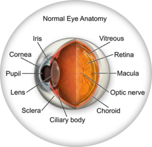
cross-section of the eye
What is accommodation?
Accommodation refers to the ability of the eye to change the shape of its lens in order to focus on objects at different distances. When you look at something up close, the lens of your eye becomes more curved to help bring the object into focus. When you look at something far away, the lens flattens out to adjust the focus accordingly.
Accommodation is controlled by the ciliary muscle, which surrounds the crystalline lens of the eye. When the ciliary muscle contracts, it changes the shape of the lens, allowing it to focus on nearby objects. When the ciliary muscle relaxes, the lens becomes flatter, enabling it to focus on distant objects.
Accommodation is an important aspect of visual function and allows us to see objects clearly at various distances. However, as we age, the ability of the eye to accommodate gradually declines, which is why many people require reading glasses, bifocals or progressives as they get older.
What to expect at an eye exam?
During an eye exam, you will be asked to sit in a special chair and look at a chart with letters or shapes of different sizes, typically a Snellen chart or a special monitor. The optometrist will then ask you to read the chart from a certain distance (20 feet or simulated 20 feet which is why you may notice mirrors in a doctors office) while covering one eye at a time with a device known as a phoropter. This helps the optometrist determine if you need corrective lenses or if you have any vision problems, such as nearsightedness (myopia) or farsightedness (hyperopia). It’s important to note the refractive exam gets more difficult as the doctor hones in on your prescription. Once you can’t tell the difference between the choices presented, the doctor will have determined your prescription. So, it is perfectly normal to not know which is better “1 or 2” at the end. I can’t tell you how many patients stress about not being able to determine the last refraction question. Fear not. “You did good!!”
The optometrist will also shine a light into your eyes using a small flashlight called a trans-illuminator. This allows the doctor to check for poor ocular mobility or nerve damage. In NJ, a doctor must examine certain features of the eye in order to qualify as a “comprehensive exam.” To get a closer look at your eye’s structure, such as the cornea, iris, and retina the doctor will use a bio-microscope called a slit lamp.
Additionally, the optometrist may perform a test called tonometry, which measures the pressure inside your eye. This is important for detecting glaucoma, a condition that can damage the optic nerve and cause peripheral vision loss.
Floaters what are they?
Eye floaters are tiny specks, spots, or cobweb-like shapes that seem to “float” across your field of vision. They can appear in one or both eyes and move around as you move your eyes. Floaters are actually tiny clumps of cells or fibers that are suspended in the vitreous, the clear gel-like substance that fills the inside of your eye.
When light enters your eye, it passes through the vitreous and is projected onto the retina, which sends visual signals to the brain. However, floaters can cast a shadow on the retina, causing you to see small spots or shapes that seem to drift across your vision.
Floaters are quite common and are usually harmless. They often occur as a natural part of the aging process, as the vitreous shrinks and becomes more liquid over time, which can cause tiny clumps of cells or fibers to become visible. Floaters can also result from other causes, such as injury to the eye, inflammation, or certain medical conditions.
While most floaters are benign and don’t require treatment, it’s important to see an eye doctor if you notice a sudden increase in the number or size of floaters, or if you experience flashes of light or a loss of peripheral vision. These could be signs of a serious condition, such as a retinal tear or detachment, which requires urgent medical attention.